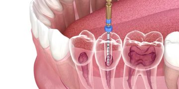
The 3D model of the root canal system of the sample presented in Fig.
Par un écrivain mystérieux
Description
Download scientific diagram | The 3D model of the root canal system of the sample presented in Fig. 2. The additional canal is marked by arrow from publication: Root Canal System Analysis with a Group of First Permanent Molars of Upper and Lower Jaw | A progressive bacteria invasion on tooth tissues leads to pulp inflammation, microabscesses of the pulp, destruction and in consequence inflammation of periapical tissues. Therefore the aim of endodontic treatment is three dimensional debridement of a root canal from the vent | Root Canal, Molar and Endodontics | ResearchGate, the professional network for scientists.
Root canal morphology and variations in mandibular second molars

Images of the crown, roots, and root canal system of a maxillary

Reliability and accuracy of dental MRI for measuring root canal
Comparison of GentleWave system and passive ultrasonic irrigation
Diagnostics, Free Full-Text

Representative 3D reconstructions of the external and internal

The C-shaped root canal systems in mandibular second molars in an

Schematic diagrams showing the root canal configuration according
:sharpen(level=0):output(format=jpeg)/up/dt/2020/06/Clinical-management-of-maxillary-1.jpg)
DT News - International - Clinical management of maxillary second

Dentistry Journal, Free Full-Text

Aetiology, incidence and morphology of the C‐shaped root canal
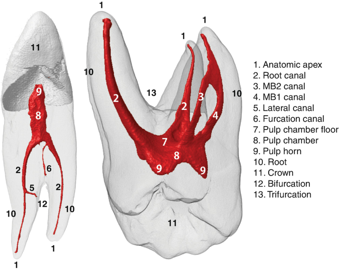
3D Visual Glossary of Terminology in Root and Root Canal Anatomy

3D models showing the external anatomy of 3 mandibular molars in

Root Canal Anatomy of Maxillary and Mandibular Teeth
depuis
par adulte (le prix varie selon la taille du groupe)