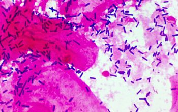
Light microscopy of Lactobacillus rhamnosus E/N (a, b) and PEN (c, d).
Par un écrivain mystérieux
Description
Download scientific diagram | Light microscopy of Lactobacillus rhamnosus E/N (a, b) and PEN (c, d). Cells suspended in PBS and mixed with ammonium sulfate 0.02 M, pH 6.8 are shown in a and c, arrows from publication: The effect of cell surface components on adhesion ability of Lactobacillus rhamnosus | The aim of this study was to analyze the cell envelope components and surface properties of two phenotypes of Lactobacillus rhamnosus isolated from the human gastrointestinal tract. The ability of the bacteria to adhere to human intestinal cells and to aggregate with other | Lactobacillus rhamnosus, Adhesion and Exopolysaccharide | ResearchGate, the professional network for scientists.

Development and Characterization of Lactobacillus rhamnosus
Lactobacillus rhamnosus

Complete genome sequence of Lactobacillus rhamnosus Pen, a

Lactobacillus rhamnosus bacteria, SEM - Stock Video Clip - K007

Zearalenone Adsorbent Based on a Lyophilized Indigenous Bacterial
Frontiers Lactobacillus rhamnosus LB1 Alleviates Enterotoxigenic
Animals, Free Full-Text
Frontiers Lactobacillus rhamnosus ameliorates acne vulgaris in

Lactobacillus rhamnosus GG Genomic and Phenotypic Stability in an
depuis
par adulte (le prix varie selon la taille du groupe)