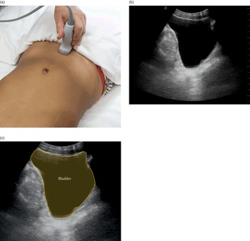
Probe position and normal images obtained during E-FAST examination.
Par un écrivain mystérieux
Description
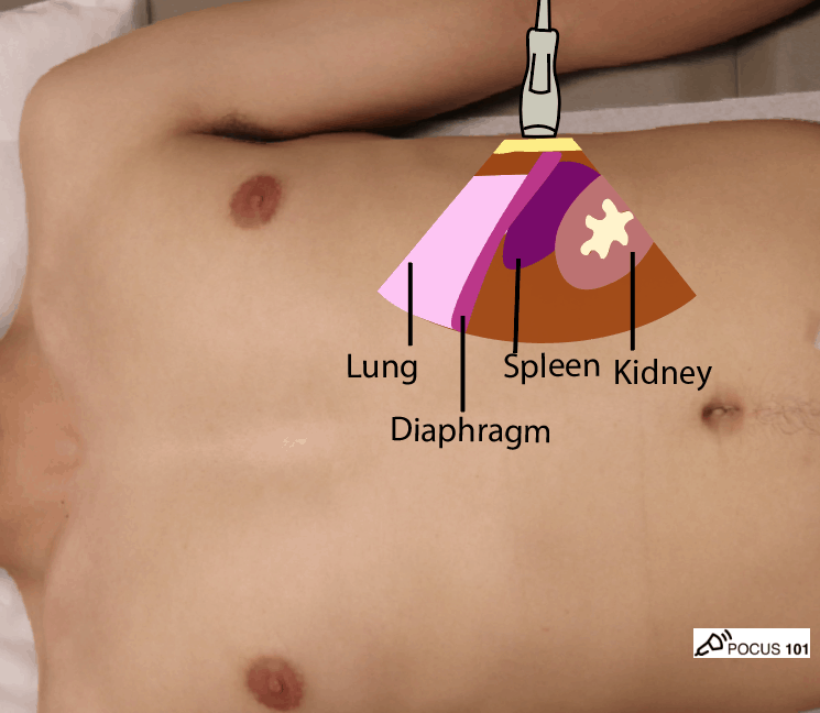
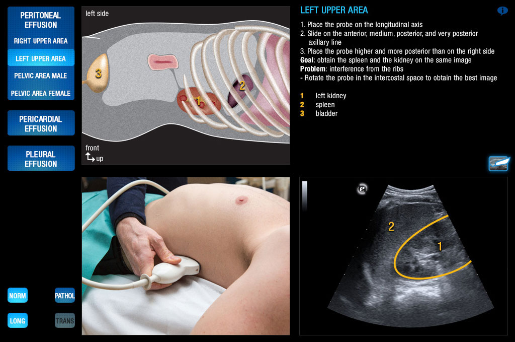
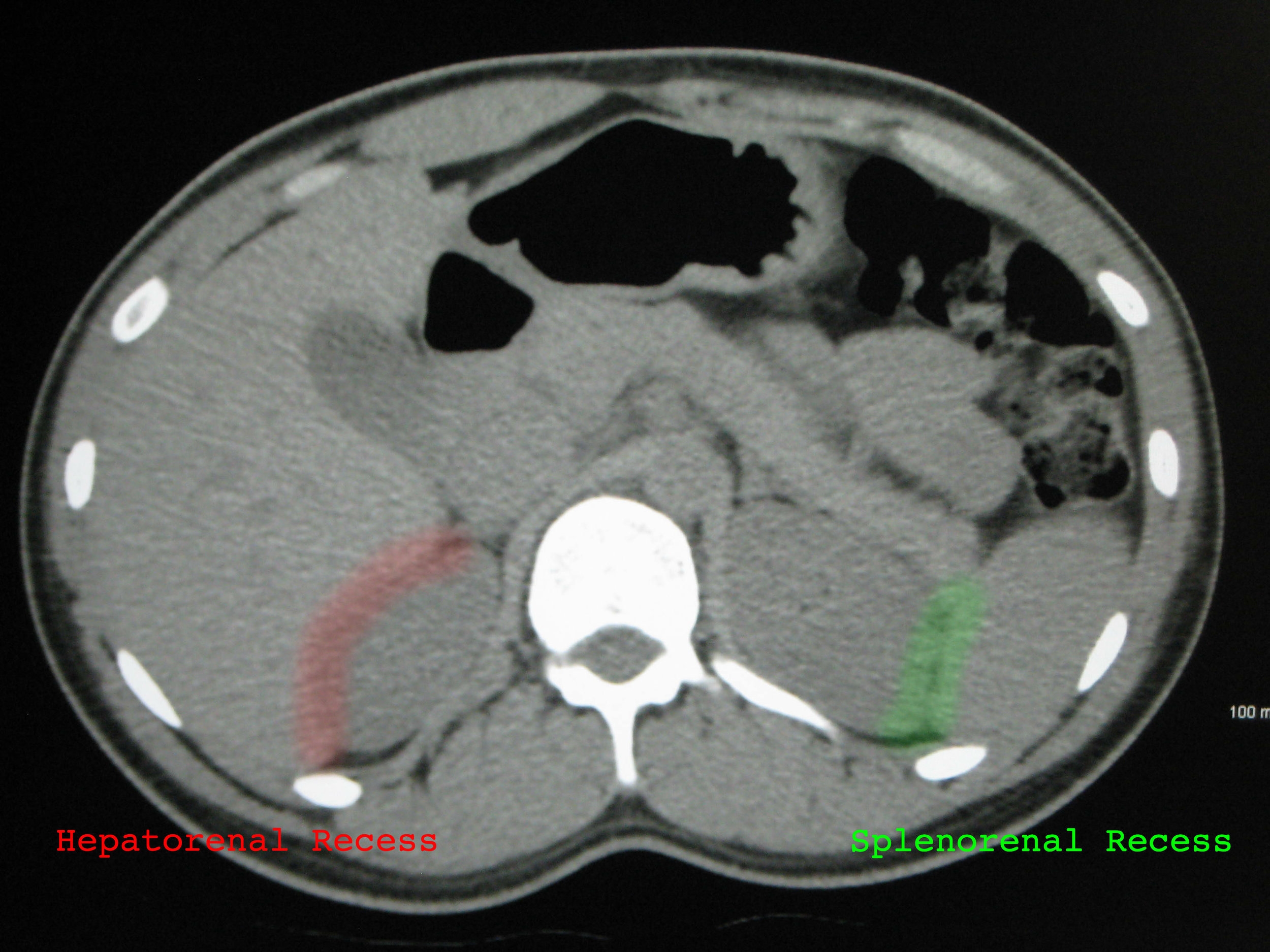
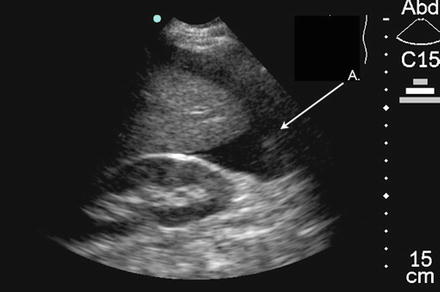
Download scientific diagram | Probe position and normal images obtained during E-FAST examination. (A) Right upper quadrant view demonstrating interface between liver and kidney (Morison's pouch). (B) Left upper quadrant view demonstrating spleen and kidney interface. (C) Left transverse view of the bladder. (D) Subcostal or subxiphoid view using the liver as a window to view the heart. (E) Anterior lung view. (F) Anterior lung view with US set to motion mode: this depicts a 1-dimensional view (marked on the top of the screen) as it changes over time (marked on the bottom of the screen); straight lines represent static soft tissue above the granular pattern representing the sliding of the pleura back and forth over time. E-FAST, Extended Focused Assessment with Sonography in Trauma examination. Used by permission from Introduction to Bedside Ultrasound, Vol 1, Dawson M, Mallin M, eds. Lexington, KY: Emergency Ultrasound Solutions; 2012: chap 1. from publication: Point-of-Care Ultrasound in Established Settings | The original and most widely accepted applications for point-of-care ultrasound (POCUS) are in the settings of trauma, shock, and bedside procedures. Trauma was the original setting for the introduction of POCUS and has been standardized under the four-plus view examination | Point-of-Care Systems, Ultrasound and Ultrasonography | ResearchGate, the professional network for scientists.
eFAST Ultrasound Exam Made Easy: Step-By-Step Guide - POCUS 101

Focused Assessment with Sonography for Trauma (FAST) scan, Radiology Reference Article
E-Fast
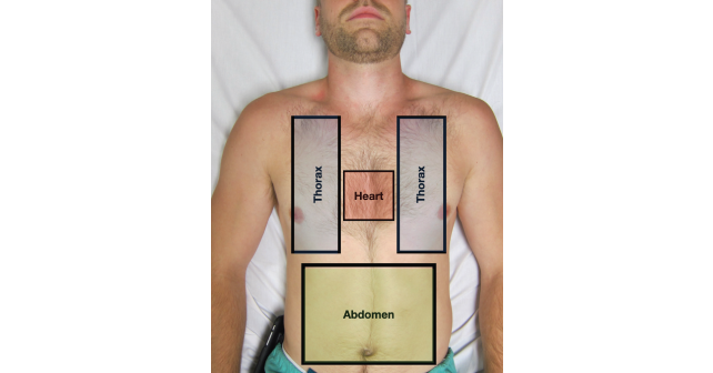
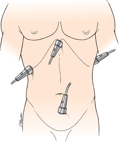
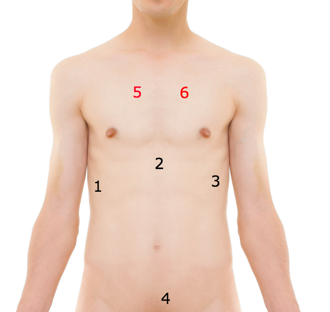
Probe placement for e-FAST: (1) subxiphoid (pericardial window); (2)
Bedside Ultrasound For Surgeons
Scanning School - FAST and eFAST — Taming the SRU

PDF] The FAST and E-FAST in 2013: trauma ultrasonography: overview, practical techniques, controversies, and new frontiers.
The extended focused assessment with sonography for trauma (E-FAST) (Chapter 4) - Pediatric Emergency Critical Care and Ultrasound
Scanning School - FAST and eFAST — Taming the SRU
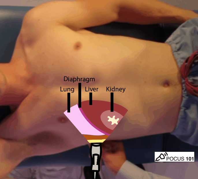
eFAST Ultrasound Exam Made Easy: Step-By-Step Guide - POCUS 101
:max_bytes(150000):strip_icc()/2782256_color1-5bc4a12fc9e77c00514acdf1.png)
Prostate Biopsy: Pain During Test, Effects, Results

Rectal examination - Wikipedia
Scanning School - FAST and eFAST — Taming the SRU
Ultrasound in Trauma Critical Care
depuis
par adulte (le prix varie selon la taille du groupe)